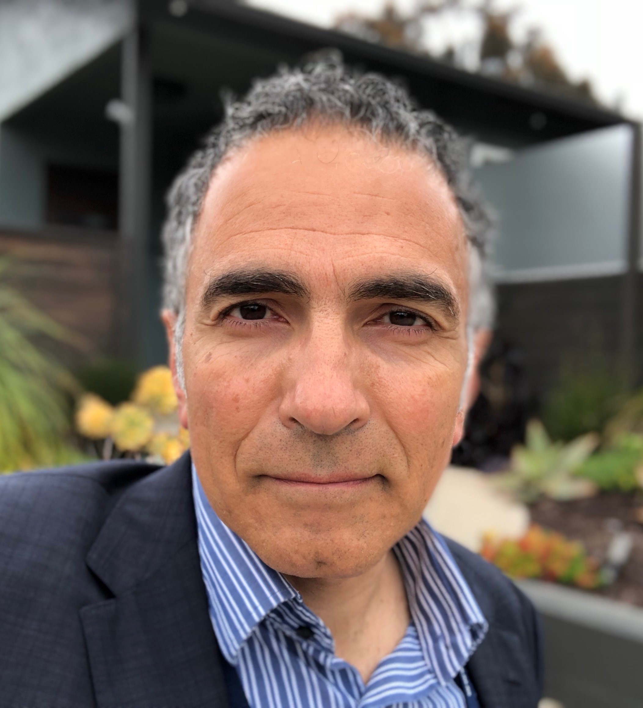
Mallar Bhattacharya, MD
School of Medicine – Medicine
Pilot for Junior Investigators in Basic and Clinical/Translational Sciences
Macrophage Function in Lung Fibrosis
Lung fibrosis is a highly morbid disease with few disease-modifying treatments and often leads to respiratory failure requiring lung transplant. A better understanding of disease mechanisms is crucial, and little is known about the function of macrophages, which have been noted to be plentiful in-patient tissue samples. This RAP award will allow me to test hypotheses motivated by our recent single cell studies with lung macrophages, where we found a unique, monocyte-derived population that drives fibrosis in mouse and human models. Our data suggest that macrophages are pro-fibrotic, and the current research aims at developing macrophage pathways for therapeutic targeting.

Peder Larson, PhD
School of Medicine - Radiology and Biomedical Imaging

Maria Roselle Abraham, MD
School of Medicine - Cardiology
Team Science Award
Hyperpolarized 13C Metabolic Imaging for Preclinical Detection of Hypertrophic Cardiomyopathy
Hypertrophic Cardiomyopathy (HCM), the most common inherited cardiac disease, has a worldwide prevalence of ~1:500, and is associated with high morbidity (heart failure, atrial fibrillation, sudden death, stroke) and mortality. However, detecting HCM prior to clinical presentations of symptoms is very challenging, even in families with known causal mutations due to incomplete penetrance and phenotypic heterogeneity. Our team plans to develop a new approach for early detection of HCM using Hyperpolarized 13C Magnetic Resonance Imaging (MRI), an emerging clinical imaging modality that uses non-toxic contrast agents with no ionizing radiation for non-invasive metabolic imaging (https://radiology.ucsf.edu/research/labs/hyperpolarized-mri-tech). This RAP award supports the creation of a new partnership between Radiology and Cardiology to pursue this goal, supporting development and testing of this unique technology. These methods can be rapidly translated for human studies using Hyperpolarized 13C MRI in order to improve care and outcomes for HCM patients and their family members.

Huinan Marcus Li, PhD
School of Medicine – Ophthalmology
Alzheimer Disease Research Center (ADRC) Developmental Projects
Defining the Role of Granulin in Astrocyte Function and Neurodegeneration
Astrocytes are the most abundant non-neuronal cell type in the brain. When specific signaling pathways are compromised, astrocytes can become reactive and can start eliminating synapses, ultimately leading to neuronal cell death during both natural aging and age-related neurodegenerative diseases. Our research aims to understand how astroglial activation is initiated in Alzheimer’s Disease and Related Diseases (ADRD). We will focus on this question by studying a gene called granulin (GRN). This gene has been linked to aging in the human cortex and loss of GRN inevitably leads to astroglial activation and a devastating neurodegenerative disease termed Frontal Temporal Lobar Degeneration (FTLD). To begin to tackle this question, we propose investigating the molecular mechanisms of GRN signaling on astroglial activation. The goal of our work is to identify the factors in astrocytes that promote brain homeostasis, aging and disease, and potentially lead to the discoveries of novel drug target in cellular level to treat neurodegenerative diseases. This award will allow me to continue my ongoing stated research, as well as helping me with my future career as an independent investigator.

Sushama Telwatte, PhD
Department of Medicine, San Francisco VA Health Care System and UCSF
Mentored Scientist Award Program in HIV/AIDS
Defining the cellular and viral factors underlying HIV latency at the single cell level
For individuals living with HIV, antiretroviral therapy is not curative and individuals experience sustained immune activation, organ damage and reduction in life expectancy. The major barrier to a cure for HIV is the establishment of latent infection of resting CD4+ T cells. Latently infected CD4+ T cells are exceedingly rare and no methods exist to distinguish latently infected from uninfected patient cells, rendering the study of HIV latency in vivo particularly challenging. The development of primary cell models of latent infection has greatly advanced our understanding of the establishment, maintenance and reactivation of HIV latency, but the precise mechanisms that underlie lifelong maintenance of latency remain unclear. This study will employ single cell technologies to probe the transcriptome of infected cells to uncover the mechanisms of HIV latent infection and reactivation. The outcomes of these studies will significantly advance our understanding of the cellular factors that contribute to the establishment and maintenance of latent vs. productive infection and could lead to new therapies to disrupt latency and/or target latently infected cells. From a personal standpoint, this award will enable me to develop key skills, cultivate productive collaborations with world-leading researchers and research institutes, and will be invaluable for my career development.

Akinyemi Oni-Orisan, PhD/PharmD
School of Pharmacy – Clinical Pharmacy
Pilot for Junior Investigators in Basic and Clinical/Translational Sciences
Development of an infrastructure to support precision medicine research for proprotein convertase subtilisin/kexin type 9 (PCSK9) inhibitor users
PCSK9 inhibitors are the first and only class of LDL-C lowering therapy found to reduce major adverse cardiovascular events on top of high-intensity background statin therapy. However, a proportion of patients treated with PCSK9 inhibitors have inadequate LDL-C reduction. PCSK9 inhibitors are expensive (~$14,300 per patient yearly); it is not feasible to use trial-and-error strategies to determine which patients will respond to therapy. The overarching goal of this project is to discover biomarkers that predict how patients will respond to PCSK9 inhibitor therapy (efficacy and safety). In collaboration with Dr. Crystal Zhou (Co-Principal Investigator of current RAP award), Dr. Eveline Stock (UCSF Cardiology), and Dr. John Kane (director of the UCSF Adult Lipid Clinic), we would ultimately like to maintain a longitudinal cohort of PCSK9 inhibitor users including robust phenotype data and biological specimens. In order to achieve this long-term research goal, the proposed pilot award will facilitate the construction of the initial cohort and collection of samples. The pilot will also conduct some preliminary exploratory analyses to characterize this population including the identification of both clinical and genetic predictors of LDL-C response to therapy. Results will be used as preliminary data for the pursuit of subsequent external funding to ensure that we can continue to characterize this exciting class of lipid-modifying therapy.

Jenise Wong, MD PhD
School of Medicine – Pediatrics, Division of Endocrinology
Pilot for Junior Investigators in Basic and Clinical/Translational Sciences
SOY-T2 - Sensors On Youth with Type 2 diabetes: A pilot and feasibility study
The prevalence of type 2 diabetes (T2D) in youth is increasing, but current standard therapy for youth with T2D is inadequate, as a large majority of teens with T2D continue to show deterioration in glycemic control, rapid disease progression, and high rates of complications. Technology such as continuous glucose monitoring (CGM) is associated with more optimal glycemic control in people with type 1 diabetes, which may be due to increased awareness of glucose profiles in response to diet, medications, or physical activity. However, research on the use of CGM systems in adolescents with T2D is limited, and few youth with T2D are prescribed CGM in clinical practice. The objective of this proposal is to investigate the feasibility of the use of CGM in youth aged 13 to 21 years with T2D in an open-label pilot randomized controlled trial (RCT). The results of this study will directly inform an NIH R-level RCT of the use of CGM in youth with T2D to improve clinical, behavioral, and patient-oriented outcomes.

Mehrdad Arjomandi, MD
UCSF Human Exposure Laboratory
Divisions of Pulmonary/Critical Care Medicine & Occupational/Environmental Medicine
Pilot for Established Investigators in Basic and Clinical/Translational Sciences
Airway Macrophage Population and Function Alteration After Ozone Exposure in COPD
Chronic obstructive pulmonary disease (COPD) is the third leading cause of death in the United States. Patients with COPD are routinely exposed to indoor and outdoor air pollution, which appears to cause escalation of their respiratory symptoms, a process called exacerbation, with resulting need to seek medical attention. This research proposes to evaluate the impact of changing populations of lung innate immune cells during the pollutant-induced airway injury using an experimental model of inhalational exposure to ambient levels of a component of air pollution (ozone) in COPD patients, and longitudinal sampling of their airway immune cells via bronchoscopy. Our overall hypothesis is that inhalational challenge to ambient levels of ozone in patients with COPD provides a safe human model to study intraluminal shifts in the population and polarization of macrophages for the study of innate immunity processes relevant to pollutant-induced COPD exacerbation, and possibly other causes of exacerbation.

Mekhail Anwar, MD, PhD
School of Medicine - Radiation Oncology
Pilot for Junior Investigators in Basic and Clinical/Translational Sciences
Intraoperative Imaging of Microscopic Residual Tumor Cells and Lymph Node Involvement Using an Ultra-Thin Molecular Imaging Skin
Microscopic tumor foci, which cannot be seen or felt, are often left behind in the tumor bed and lymph nodes during cancer surgery, increasing cancer recurrence and metastases, respectively. Despite the advent of molecularly targeted imaging agents, image guided cancer surgeries remain hindered by our ability to visualize labeled cells intraoperatively by the imagers themselves. This RAP project unlocks the power of new, inorganic optical nanoparticles to significantly advance in vivo intraoperative molecular imaging for cancer by developing an ultra-thin molecular imaging skin, just <200 μms thin, made from computer-chip technology, integrated on virtually any surgical instrument, for real-time, single-cell visualization of tumor cells intraoperatively.
While applicable to all cancer types, success of this project will aid in solving the problem of tumor cells left behind in both breast and prostate cancer surgeries. For example, breast cancer 1 in 4 women have tumor cells left behind and require a reoperation - estimated to be 37,000 operations per year. In prostate cancer, upwards of 25% of advanced prostate cancers can have tumor cells left behind. Precision mapping of all tumor cells intraoperatively will not only help surgeons remove all tumor cells, but will enable radiation oncologists to be more precision and effective when delivering post-operative radiation. This award is critical to advance this innovative, out-of-the box concept for further funding and clinical translation.